Imaging Flow Cytometry
Imaging flow cytometry is a powerful technique for high-throughput single-cell analysis. It provides comprehensive analysis and in-depth imagery of every individual cell and is used in applications such as cancer screening.
Blood cells are aligned in a microfluidic channel and scanned by an ultrafast pulsed laser beam, and the output is captured by high-performance digitizers via photomultiplier tubes (PMTs). Both the flow speed and image quality are superior to conventional CMOS-based approaches, and due to the high throughput single-cell analysis and characterization enable high statistical accuracy by millions of captured cell images.
- High dynamic range is required to achieve superior image quality
- High sampling rate enables high microfluidic channel flow rate and system-level throughput
- Open FPGA is crucial for real-time image pre-processing
- High data transfer rate is needed in order to support high-speed image post-processing and data storage
- Peer-to-peer GPU streaming offers additional benefits for cell characterization
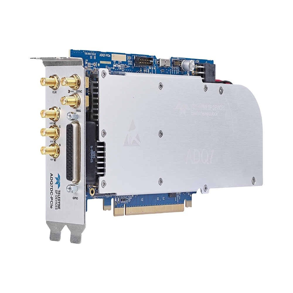
Our imaging flow cytometry customers typically use ADQ7DC in PCIe form factor. It is a single-/dual-channel, 14-bit digitizer with up to 10 GSPS sampling rate and it features a large on-board Xilinx Kintex Ultrascale FPGA which is open to the user.
Furthermore, ADQ7DC supports GPU peer-to-peer streaming at rates up to 6.8 GByte/s across a PCIe Gen3 x8 bus. Optional firmware and development kits can be purchased separately at any time and upgrades can be done in place without any need for system disassembly or significant downtime.